
Podiatry Foot Unit
At the Foot Unit of Clínica Premium, together with the team from the Nogales Foot Unit, we treat foot pathologies with a biomechanical approach.
Premium Foot Unit
We offer Advanced diagnostics, 3D scanning and personalized treatments. Experts in Minimally invasive open-field foot surgery.
Foot Unit Nogales and Podoactiva.
Podoactiva, [Company Name], a leader in podiatry and biomechanics, is present in more than 150 clinics in Spain and several other countries, including [Country Name]. Nogales Foot Unit. Its exclusive technology 3D Scan Sport Podoactiva®, patented worldwide, allows design high-precision custom templates, already used by more than 350,000 patients.
In Podoactiva, Clinical excellence, technological innovation, and R&D are fundamental pillars. Their patented system 3D Scan Sport Podoactiva® It allows for a high-precision foot scan to create custom and robotic templates, tailored to each patient. More than 350,000 people, including 3,500 elite athletes, already rely on this technology, also available in the Nogales Foot Unit, which shares the same philosophy of continuous improvement and cutting-edge care.


Foot surgery – Minimally invasive surgery
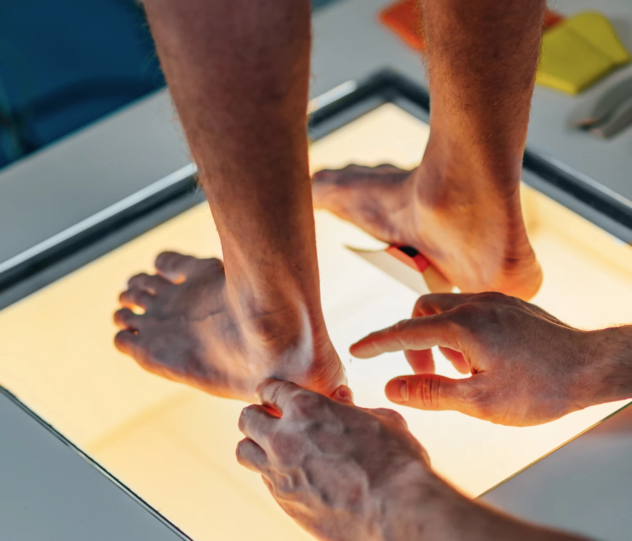
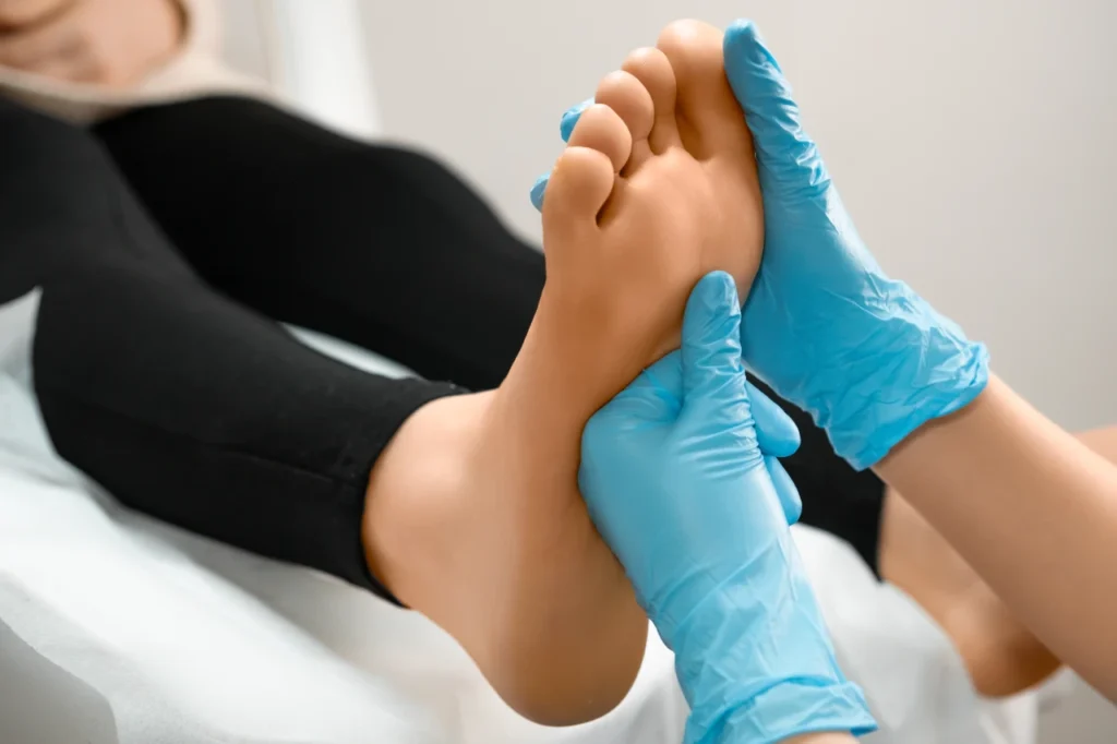
During the patient's initial visit, their condition is assessed to arrive at the correct diagnosis. We then explore possible treatment options and evaluate each one until we find the most appropriate solution. For this reason, we utilize state-of-the-art equipment that allows us to establish an accurate diagnosis.
In this unit we offer a wide range of treatment options for our patients, trying to find a definitive solution for them.
Dr. Nogales has been a member and professor of the Spanish Association of Podiatric Surgery (AECP) since 2010 and a member and instructor of the Spanish Association of Minimally Invasive Surgery (AEMIS). He has over 10 years of experience in this field, both as a teacher and researcher.
Major surgery operating room
Our facilities include an outpatient surgery operating room for performing foot procedures such as bunion removal, Morton's neuroma treatment, claw toe correction, hindfoot surgery, minimally invasive surgery, ultrasound-guided procedures, and tumor pathology, among others. We have a team of anesthesiologists to perform procedures with local anesthesia and sedation for your comfort. We also offer a private room with a bathroom equipped for patient rest after surgery, allowing you to return home the same day once fully recovered.
Minor surgery operating room
In addition to this major surgery suite, we have a minor surgery suite for procedures performed under local or regional anesthesia to treat warts, cysts, and nail issues. This team is specialized and highly regarded in osteoarticular surgery, including treatments for hallux valgus (bunions), tailor's bunions, claw toes, metatarsalgia, heel spurs, exostoses and bone deformities, and flat feet in children and adults.
Functionality and aesthetics
Given our training, we believe that minimally invasive surgery and open surgery are perfectly compatible, so much so that we usually combine both techniques in most of our patients, thus obtaining a much more functional and aesthetic result.
Biomechanics – Gait analysis – Sports podiatry
Our working system allows us, based on a personalized gait analysis, to scan the shape of your foot in 3D and send our treatments to a team that integrates Engineers and Podiatrists to design a unique, 3D printed and effective treatment to solve your pathology or prevent injuries.
How do we perform the treatment?
- We decide on the treatment based on a biomechanical examination or study of the gait of our patients with the aim of preventing injuries or treating any foot pathology.
- After the biomechanical exploration, a 3D image of our patients' feet is obtained using a scanner and sent to headquarters so that engineers can design the treatment, print it, and send it back to the clinic.
- This process usually takes 15 days.
- It is possible to track orders to know their status.
Result
A 3D printed insole, the most advanced on the market, which provides the necessary resistance to avoid pathological situations while also presenting a degree of flexibility necessary to allow the natural movement of the foot.
Suitable for the general population, children and athletes.
Interventional ultrasound
Ultrasound is a diagnostic imaging procedure with numerous advantages. It is a fast, non-invasive technique that does not emit ionizing radiation. Thanks to ultrasound, certain types of procedures can be performed, including radiofrequency ablation.
Radiofrequency ablation works by interrupting nerve conduction, preventing the nerve from transmitting pain signals. This technique resolves the problem in 90% cases.
This procedure is performed in a single session, with local anesthesia in the foot. The patient can walk out that same day and can resume their normal life almost immediately.
Radiofrequency for the treatment of Morton's neuroma
Morton's neuroma is defined as a compressive neuropathy, which consists of a degeneration of the plantar digital nerve (which runs between the toes) accompanied by fibrosis or thickening around the nerve.
It is usually located between the 3rd and 4th metatarsals (in 66% cases), primarily due to two reasons: these are the metatarsals that move the most during gait, and the nerve located in this space is anatomically thicker than the other digital branches. It can also be found between the 2nd and 3rd metatarsals in 32% cases (Hauser's neuroma) and between the 4th and 5th metatarsals in 6% cases (Iselin's neuroma). It is uncommon for it to appear in more than one of these locations simultaneously.
Morton's neuroma is a fairly common pathology seen in consultation, being more frequent in women than in men in a 4:1 ratio. It is usually bilateral in 21% of cases.
The most common symptom is a burning pain in the sole of the foot, between the metatarsal heads. This pain usually radiates to the two corresponding toes. The patient typically experiences a sharp pain that continues along the sole or top of the foot and worsens with tight shoes or high heels.
The diagnosis is primarily clinical and ultrasound-based; therefore, we have state-of-the-art equipment that allows us to visualize the neuroma, determine its size, and establish the most appropriate treatment options for each case.
Pediatric podiatry
Why is it important to care for the feet of young children?
At Podoactiva we are specialists in pediatric podiatry and have specific protocols for the diagnosis and pathologies of the foot and gait in children.
It is really important to keep track of the evolution of children's gait, because detecting a pathology in time can make the difference between applying a treatment to solve that problem at its root or not.
If the appropriate diagnosis and treatment are carried out early, most foot and gait problems can be solved through simple treatments such as adapted exercises, custom insoles and splints, among others.
Even if no abnormality is detected in the child's gait, it's important to perform a biomechanical gait analysis. Children's feet are constantly evolving and are very flexible; they may not report any pain at first, but it's best to ensure the child's gait is correct and to schedule regular checkups.
Regenerative medicine
Among the different regenerative therapies we find Plasma Rich in Growth Factors, also known as Platelet Rich Plasma (PRP).
These platelets carry growth factors, proteins that play a fundamental role in the regeneration of damaged tissue. The aim of this therapy is therefore to accelerate the healing and tissue repair process, in addition to its anti-inflammatory effect.
The treatment involves autologous plasma infiltration; that is, blood is first drawn from the patient and centrifuged to obtain plasma with a platelet concentration much higher than that normally found in the bloodstream. This plasma is then injected under ultrasound guidance into the affected area under local anesthesia.
This procedure has many indications, including:
- Plantar fasciitis: PRP accelerates the healing of the fascia, reduces inflammation, improves elasticity, and thus achieves a reduction in pain.
- Chronic tendinopathies and ligament injuries.
- Muscle injuries.
- Degenerative joint injuries, such as osteoarthritis of the big toe or "Hallux Rigidus," where there is damage to the joint due to wear and tear of the articular surfaces, can be treated with PRP injections. These injections improve mobility and reduce pain and inflammation.
- For bone fractures and stress fractures, PRP achieves rapid repair due to its direct infiltration into the bone lesion thanks to interventional fluoroscopy control.
Diabetic foot
Dr. Zumaquero and Dr. Nogués are in charge of the diabetic foot unit. Diabetes mellitus is a disease with an increasingly significant prevalence in our Western society. Foot involvement is one of the most frequent and feared complications, giving rise to what we commonly know as "diabetic foot.".
Due to high blood glucose (sugar) levels, a number of alterations occur related to decreased blood circulation and loss or reduction of sensation. These factors can predispose individuals to conditions ranging from the development of plantar ulcers to the fatal complication of amputation. The vast majority of amputations could be avoided by prioritizing prevention and adopting healthy lifestyle habits.
Some tips to avoid possible complications are:
- Inspect your feet daily to check for any injuries or redness.
- Pay special attention to hygiene; it is important to wash your feet every day with warm water and soap, checking the temperature beforehand to avoid burns.
- Dry them thoroughly, moisturize them, and use powder in cases of excessive sweating.
- Cut your nails straight across, avoiding leaving any corners that could cause injury.
- Wear wide, comfortable shoes. Check the inside of your shoes daily for anything that could hurt your feet.
In addition to these tips, a visit to the Podiatrist is advisable to carry out a neuropathic and vascular study in order to determine the degree of impairment, as well as a biomechanical study with the aim of eliminating areas of hyperpressure that may predispose to the appearance of ulcers through insoles.
Common pathologies
Foot care and health are fundamental to maintaining a good quality of life, mobility, and independence in our daily lives. By continuously supporting the body's weight and absorbing the impact of each step, the structures of the foot are exposed to various biomechanical imbalances, continuous pressure, genetic factors, and the use of inappropriate footwear.
HAV (bunion) surgery
Hallux abductus valgus, or what is commonly known as a bunion, is a bony deformity of the metatarsophalangeal joint of the big toe, causing it to deviate toward the other toes. It is characterized by the formation of a bony bump, the main symptom of which is pain that worsens when walking. Inflammation and redness may also appear in the area due to friction with footwear. It is more common in women due to the use of high heels and shoes that are too narrow.
What are the causes?
There is no single exact cause that determines its appearance, but there are a number of predisposing factors:
- Biomechanical alterations: ligamentous hyperlaxity, hypermobility of the first ray…
- Inflammatory joint diseases.
- Neuromuscular diseases.
- Footwear.
- Bone morphological alterations.
- Trauma.
- Iatrogenesis.
- Genetic factors.
What are the treatment options?
Treatment options will depend on the degree of deformity and pain. In cases where conservative treatment does not relieve symptoms, surgery is considered.
There are many surgical techniques described for the treatment of this deformity that can be performed through minimally invasive surgery, erroneously known as "laser surgery" since no type of laser is used but the correction of the deformity is carried out through millimeter incisions.
The other approach option is through open surgery techniques where the incision is somewhat more extensive but is equally aesthetic, avoiding scars thanks to closure with intradermal sutures.
There is no single best technique; it all depends on the patient's characteristics and the professional's assessment, who will determine the best option for each individual case.
Hallux Rigidus
Hallux rigidus is a condition characterized by reduced dorsiflexion (upward bending) of the metatarsophalangeal joint of the big toe, accompanied by mechanical pain. Some patients even experience stiffness and an inability to perform the movement.
It is more common in men between the ages of 40 and 60. There are several causes, including an excessive length of the first metatarsal, rheumatic diseases (gout), and repetitive trauma.
Degenerative changes occur at the cartilage level that lead to early osteoarthritis, visible by ultrasound and through fluoroscopy.
Prior to hallux rigidus, we find the stage of "functional hallux limitus," where movement limitations and discomfort are less severe. It is at this stage that progression to hallux rigidus should be halted with conservative treatment using custom orthotics to improve biomechanics, and injections of PRP, hyaluronic acid, and collagen.
When conservative treatments are ineffective, surgery is used to solve the problem permanently.
Metatarsalgia
Metatarsalgia is pain that occurs in the front and sole of the foot, where the heads of the five metatarsals are located. These bones support the weight of the entire body when the foot pushes off forward during a step, a moment when they receive excessive pressure. This repetitive pressure generates inflammation that eventually causes a sharp, burning pain, the appearance of hyperkeratosis (calluses) in the area, and even joint displacement in the worst cases.
The main cause is poor pressure distribution, but other factors also play a role, such as being overweight, digital deformities, a high arch, or wearing inappropriate footwear such as excessively high heels.
The treatment will depend on the cause of the problem, ranging from weight loss or the creation of custom-made insoles that allow for better distribution of loads to eliminate pressure points, to surgical treatment if conservative treatment fails.
Claw fingers
Claw toe is a deformity that occurs in the toes due to a muscular and tendon imbalance. There are two types of claw toe:
- Flexible claw: the deformity corrects itself by extending the finger.
- Rigid claw: it is impossible to correct the deformity by extending the finger.
The causes of this condition can be congenital, neurological, muscular, articular, or due to biomechanical alterations. High arches, flat feet, or bunions are examples of conditions that predispose individuals to developing them.
A large majority of patients suffering from this condition wear inappropriate footwear with narrow toes or heels, so the type of footwear greatly influences the development and evolution of the deformity.
What are the signs and symptoms?
Apart from the deformity itself, there are a number of signs and symptoms specific to this pathology, such as:
- Appearance of helomas (calluses) on the fingertip and back due to friction and rubbing with footwear.
- Pain when walking that intensifies when doing sports.
- Inflammation of the joint.
What are the treatment options?
In cases where conservative treatment has failed or the deformity is in an advanced stage, surgery is considered as a definitive solution.
Interdigital corns (Corns)
An interdigital heloma, or what is commonly known as a corn, is a callus located between the toes, most frequently between the fourth and fifth toes. It originates from pressure and friction between the toe bones, which can be observed with fluoroscopy.
It produces a sharp pain that is aggravated by the use of narrow-toed shoes.
How to solve the problem?
There are different types of solutions for this condition. The temporary solution consists of the non-invasive removal of the corn periodically, and to reduce friction, a custom-made silicone orthotic can be used, along with appropriate footwear. However, if a permanent solution is desired, we resort to minimally invasive surgery to smooth down the bony protrusion causing the pain. It is a simple and quick procedure performed through a tiny incision.
Plantar fasciitis
Plantar fasciitis is an inflammation of the plantar fascia, a band of elastic tissue that runs from the calcaneus (bone located in the heel) to the metatarsal area (under the toes).
It is characterized by intense morning pain that decreases throughout the day and with rest. It is a fairly common condition in athletes and runners. However, it can also occur in people who do not participate in sports. Studies indicate that 10% of the population may experience it at some point in their lives.
There is a direct relationship between plantar fasciitis and shortening of the posterior leg muscles, and it is common in women who continuously wear high heels.
Regarding gait and foot type, there is a greater possibility of suffering from this pathology in cavus feet, valgus feet and feet with a tendency to pronation due to an increase in tension from a decrease in support.
We offer multiple treatments for this condition, which will vary depending on the duration of the condition. In the acute phase, it can be managed with anti-inflammatory medications and physical therapy exercises, as well as a gait analysis to assess whether there is a connection to the patient's walking pattern. If a connection is found, custom orthotics are recommended to manage the problem.
Regenerative medicine can also be used through the infiltration of plasma rich in growth factors (PRP) that accelerates healing and tissue regeneration.
When plantar fasciitis has been present for a long time and conservative treatment has failed, there are still solutions thanks to recent advances in surgery. One option is interventional ultrasound, which allows for the ultrasound-guided release of the fascia through minimally invasive surgery. Another option is to perform the procedure using percutaneous or minimally invasive surgery with fluoroscopic guidance.
Tarsal tunnel entrapments
The tarsal tunnel is located on the inner side of the ankle. Tarsal tunnel syndrome is a compressive neuropathy of the posterior tibial nerve or some of its branches as it passes through this area, just below a ligament called the flexor retinaculum.
This is a very characteristic pain, described as cramping, burning, and even numbness, which may or may not radiate to the sole of the foot. It increases when walking and can intensify at night, decreasing with rest and removal of the shoe.
The causes are varied: inflammatory processes or alterations that compress the nerve; traumatic injuries such as fractures, sprains, tarsal coalitions; systemic diseases; deformities such as flat feet or valgus that cause excessive tension, with the consequent compression of the nerve.
Diagnosis relies heavily on a thorough medical history and physical examination, including a series of tests and maneuvers, as well as complementary imaging such as magnetic resonance imaging (MRI). Another diagnostic approach involves selective, ultrasound-guided nerve block using local anesthetic infiltration to determine which branch of the tibial nerve is compressed. We also utilize an intracompartmental pressure monitor to aid in diagnosis.
Regarding treatment, the first approach will be conservative, including custom orthotics to relieve tension, ultrasound-guided injections, and physical therapy. If the problem does not improve, surgery will be considered to release the damaged nerve branches.
Baxter's neuropathy
Baxter's neuropathy is an entrapment of the first calcaneal branch, or Baxter's nerve, which can be caused by biomechanical factors (such as gait abnormalities) or direct compression from a space-occupying lesion. It is characterized by pain in the medial plantar area of the heel, accounting for up to 20% of cases with this condition, and is easily confused with plantar fasciitis.
Diagnosis can be complex; ultrasound-guided infiltration with local anesthetic is of great interest for treatment or measurement of intracompartmental pressure.
Among the treatments that can be carried out are infiltrations or radiofrequency thanks to interventional ultrasound and in those cases where the above techniques do not work, surgical treatment would be indicated with the aim of decompressing the nerve.
Furthermore, this team performs decompression of this nerve through both minimally invasive surgery and open surgery, as required by the case.
Onychocryptosis (Ingrown toenails)
Onychocryptosis, or ingrown toenail, is a condition in which the nail grows into the skin of the toe on its lateral and/or medial edge. It most often affects the big toe, although it can occur in any toe. The ingrown nail causes pain, inflammation, and redness, and in the worst cases, can even lead to infection.
Its appearance may be due to congenital factors such as the shape of the nail, alterations in the distal phalanx,…or to triggering factors, among which are:
- Trauma.
- Repeated microtrauma due to the use of shoes with a narrow toe box and/or heel.
- Bad nail trimming.
- Biomechanics.
How to prevent ingrown toenails?
Proper nail trimming is essential, as is choosing the right footwear and appropriate running shoes for athletes. Consulting a professional in these cases is the best way to prevent future complications.
What are the treatment options?
Regarding treatment, we can find a temporary solution or a permanent solution.
Conservative treatment, or a temporary solution, involves removing the ingrown nail spicule. In some cases, the problem doesn't recur, and this treatment resolves it completely. However, this is rare, so the patient will need to see a professional periodically as the nail grows back to have the procedure repeated.
Surgical treatment, or the definitive solution, is the best option in most cases. It consists of a minor outpatient procedure performed in the office under local anesthesia of the affected finger. Generally, a partial matricectomy is performed, removing the matrix cells. The expected result is a nail where the treated edge does not grow back, with a functional and aesthetic appearance.